Current Issue

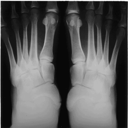
Young man with left foot pain

Chin-Hwee LEE1 and Wilfred CG PEH2
1Faculty of Medicine, National University of Singapore and 2Singapore Health Services, Singapore


Case history


A 23-year-old man was referred for pain in his left foot of 2 weeks duration following a sprain. Examination revealed vague tenderness over the medial aspect of the left mid-foot. An anteroposterior radiograph of both feet was obtained.

Q1. What are the radiological findings?

Q2. What is the diagnosis?

Q3. What is the significance of this condition?

Q4. Could an avulsion fracture give this radiology appearance?

Q5. Could a March fracture give this radiology appearance?

References

1 Schmidt H, Freyschmidt J. Borderlands of Normal and Early Pathologic Findings in Skeletal Radiology, 4th edn. New York: Thieme Medical Publishers; 1993: 815-26.

2 Apley AG, Solomon L. Concise System of Orthopaedics and Fractures, 2nd edn. Oxford: Butterworth-Heinemann; 1994: 222, 343.

Figure 1: Anertior-posterior radiograph shows a left accessory navicular bone depicted as a well-corticated rounded bone (black arrow) located dorsomedially to the normal left navicular bone (broad open arrow). Right foot is normal.