
RADIOLOGY SERIES
Lady with painless breast lump
Neerajana DODA,1 Wilfred C. G. PEH2
1Department of Diagnostic Radiology, Changi General Hospital and 2Singapore Health Services, Singapore
Case historyLady with painless breast lump
Neerajana DODA,1 Wilfred C. G. PEH2
1Department of Diagnostic Radiology, Changi General Hospital and 2Singapore Health Services, Singapore
A 52-year-old Chinese woman incidentally noticed that she had a painless left breast lump. A mammogram was obtained.
Q1 What are the mammographical findings and the diagnosis?
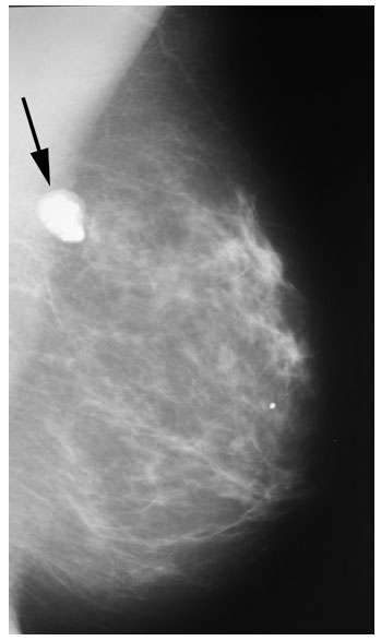
The mammogram shows a dense, lobulated, well-defined lesion in the upper half of the breast, with popcorn-like calcification (Fig. 1). There is no associated architectural distortion or microcalcification. Appearances are typical of an involuting calcified fibroadenoma.
Q2 What is a fibroadenoma?
A fibroadenoma is the most common benign solid lesion of the breast. It is produced by an overgrowth of stromal connective tissue of the lobule, often accompanied by a variable overgrowth of epithelial structures.1 The cause is unknown. They are most common in young women in their twenties and thirties, and may regress after menopause.1,2 There is no racial predilection.2 Clinically, these lesions may or may not be palpable. When palpable, they may be felt as oval, freely-mobile, rubbery masses.2 They can be hard on palpation when calcified. Being mobile on palpation, they are sometimes called breast mice. Postmenopausally, they may regress and often calcify.
Q3 What are the typical radiological findings of a fibroadenoma?
On mammography, fibroadenomas are usually seen as a well-defined, sharply-circumscribed lesion, and may be lobulated. They may have a thin lucent halo around them.1 Calcifications are common, especially in postmenopausal women. Most of the calcifications are coarse and the so-called popcorn calcifications are pathognomonic for fibroadenoma. On ultrasonography, fibroadenomas can be extremely variable in echogenic characteristics. Typically, however, they tend to be well-circumscribed, homogeneous, oval, hypoechoic lesions with sharp margins which may be lobulated and show variable acoustic enhancement.1,2 When calcified, they show extensive after-shadowing.
Q4 What is the prognosis and risk of carcinoma?
The prognosis of fibroadenoma is excellent. They are benign lesions and are not considered to have malignant potential. However, as they contain epithelium, a risk of malignancy exists. The risk of a breast carcinoma occurring within a fibroadenoma is about 3%. This is twice as likely in women who have undergone previous excision of fibroadenoma and when they are associated with cysts, sclerosing adenosis or calcifications.2
Q5 What is the treatment?
For young women who have typical features of fibroadenoma, no intervention is needed.3 Alternatively, a needle biopsy may be performed and if proven to be a fibroadenoma, it may be left alone or removed, depending on the patient. When left alone, they should be periodically monitored by physical examination and imaging, following the recommendations of the doctor. In event of change in size or development of suspicious microcalcifications, a biopsy may be required.2,3
References
1. Kopans DB. Breast Imaging. 1st edn. Philadelphia: JB Lipincott Company; 1989: 2625.
2. Roubidoux MA. Breast Fibroadenoma. http://www.emedicine.com/radio/topic109.htm
3. http://www.medicineonline.com/references/Womens_Health/ Breast_Health/Breast_Cancer/Fibroadenoma_breast/
Fig 1 Mammogram (medial lateral oblique view) of the left breast shows a dense well-defined lesion with the typical popcorn calcification (arrow) of an involuting fibroadenoma. It has lobulated margins with a faint lucent halo.
^top