
Asia Pacific Journal of Family Medicine Volume 5 Issue 3
RADIOLOGY SERIES
Woman with possible right toe fracture
Neerajana DODA1 and Wilfred C. G. PEH2
1Department of Diagnostic Radiology, Changi General Hospital and 2Singapore Health Services, Singapore.
Correspondence: Professor Wilfred CG Peh, Senior Consultant Radiologist, Programme Office (Graduate Medical School), Singapore Health Services, 7 Hospital Drive, #0209, Singapore 169611
Accepted for publication 2006.
Introduction
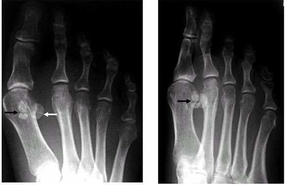
A 50-year-old woman presented with history of trauma and pain over the big toe of the right foot. Antero-posterior and oblique radiographs of the right foot were obtained
What are the radiographical findings?
There are three small bones seen in the region of the head of the metatarsal of the right big toe. Two are located medially and one laterally. The two small medial bones lie close to each other but are clearly separated from one another. They are both well-corticated. Both medial bones have well-defined margins, with the apposing surface of the distal sesamoid bone having a rounded margin and that of the proximal sesamoid bone having a slightly irregular margin (Fig. 1a, b). No obvious fracture is seen.
What is the diagnosis?
Bipartite medial sesamoid. These are the sesamoid bones of the big toe and are a normal variant. Normally, there is one medial (tibial) and one lateral (fibular) sesamoid. However, in this case, there is a bipartite medial sesamoid and a single lateral sesamoid. A bipartite sesamoid may be confused with a fracture in the sesamoid bone itself. It is therefore important to know the difference between the two entities, as a bipartite sesamoid is a normal variant.
What is a sesamoid bone?
In the big toe, sesamoid bones are seed-shaped bones that are found at the first metatarsal head. They are plantarly located within the tendon of the flexor hallucis brevis.1,2 There are usually two bones, one medial (tibial sesamoid) and one lateral (fibular sesamoid). They play an important role in the mechanics of the foot: increasing the mechanical advantage of the flexor hallucis brevis, assisting with weight-bearing under the big toe metatarsal, and they also help in elevating the metatarsal head off the ground.1,2 The medial sesamoid is usually larger than the lateral sesamoid, and plays a more important role in weight-bearing.
What is a bipartite sesamoid?
When a sesamoid develops from two ossification centres that do not fuse at maturity, it is referred to as a bipartite sesamoid. About 10% of people have this condition and in those who have it, there is a 25% chance of being bilateral.3 This condition is much more common in the medial sesamoid than the lateral sesamoid. Sometimes there may be more than two centres of ossification. The sesamoid is then called a multipartite sesamoid.
How can a bipartite sesamoid be differentiated from a fracture of the sesamoid?
First, the clinical history and site of pain should be correlated with the radiographical findings. A sesamoid fracture usually presents with an acute onset of pain and tenderness on direct palpation over the injured sesamoid. Radiographically, a fracture of the sesamoid shows sharp edges and corners, and the fracture line is almost always transverse in orientation. The well-defined cortical outline and rounded margins of a bipartite sesamoid are not present. However, because of the small size of the bones, diagnosis may be difficult, in which case a radiograph of the other foot may be useful to look for a contralateral bilateral sesamoid. Increased uptake on bone scintiscans has shown to be useful in diagnosis, if the diagnosis is still unclear on radiographs. Bone scintiscans will be positive in a fracture and negative in a bipartite sesamoid.3,4
What are the other conditions which may cause sesamoid pain?
The sesamoids may be injured or become painful due to: sesamoid fractures (as described above); sesamoiditis (inflammation of the sesamoid); sesamoid stress fracture (a break that develops slowly over a period of time); and rarely, avascular necrosis and dislocations of the sesamoid.4
References
1 . http://www.wheelessonline.com/ortho/sesamoid_bones_of_the_foot
2 . http://www.thenaturalfoot.com/sesamoid.html
3 . http://www.cpma.org/newsltr/fall2001.html
4 . http://www.med.umich.edu/1libr/sma/sma_sesamoid_sma.htm
Figure 1 - (a) Antero-posterior and (b) oblique radiographs of the right forefoot show two small sesamoid bones (bipartite sesamoid) located medially at the metatarsal head of the big toe, and one sesamoid located at the lateral aspect (white arrow). Note the sharp demarcation between the well-defined cortical surfaces of the bipartite medial sesamoids (black arrow). These are features of a normal variant rather than a fracture.
(a) (b)
^top